Introduction
Mifepristone, commonly known as RU-486, stands as a pivotal medication in early pregnancy termination. Its mechanism of action revolves around disrupting the crucial hormonal balance required for pregnancy maintenance, particularly by impeding the effects of progesterone, a hormone vital for the sustenance of the uterine environment. Understanding how mifepristone intervenes in the progesterone pathways sheds light on its capacity to detach the embryo from the uterine wall, leading to the cessation of pregnancy progression.
Understanding Pregnancy and Progesterone
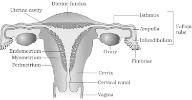
Pregnancy initiation begins with the fertilization of an egg by sperm, resulting in the formation of a zygote. The zygote undergoes several divisions and becomes a blastocyst, which eventually implants itself into the lining of the uterus, a process known as implantation. For the pregnancy to progress successfully, the blastocyst needs to attach firmly to the uterine wall and receive nutrients and support from the mother’s body. Progesterone, a hormone primarily produced by the corpus luteum (a temporary endocrine structure formed in the ovary after ovulation), plays a crucial role in maintaining the uterine lining during the early stages of pregnancy. It supports the thickening of the endometrium (uterine lining) and prevents its shedding, providing a suitable environment for the developing embryo.
Mechanism of Action of Mifepristone
Mifepristone is a synthetic steroid compound that acts as a progesterone receptor antagonist. Its mechanism involves binding to progesterone receptors in the uterus and competitively inhibiting the binding of endogenous progesterone. This interference disrupts the normal progesterone signaling pathways, leading to a cascade of effects that result in the detachment of the embryo from the uterine wall and subsequent termination of pregnancy.

Effects of Mifepristone on the Uterine Lining
Blocking Progesterone Receptors: By binding to progesterone receptors, mifepristone prevents the natural progesterone hormone from exerting its effects. Progesterone, usually responsible for maintaining the thickness and stability of the endometrial lining, becomes ineffective due to this antagonist action.
Disruption of Uterine Environment: The blockade of progesterone receptors alters the usual hormonal balance necessary for sustaining pregnancy. This disruption weakens the attachment between the developing embryo and the uterine wall.
Decreased Blood Supply: Mifepristone also causes a reduction in the blood supply to the developing placenta by affecting the growth and functioning of the trophoblast (tissue that forms the placenta). This decreased blood flow contributes to the detachment of the embryo from the uterine wall.
Induction of Detachment and Termination
As a consequence of mifepristone’s actions on the progesterone receptors and the subsequent changes in the uterine environment, the attachment between the embryo and the uterine wall becomes compromised. This detachment prevents the embryo from receiving the necessary nutrients and support for its continued growth and development. Following mifepristone administration, a second medication called misoprostol is often given to induce uterine contractions, further aiding in the expulsion of the embryo and the contents of the uterus.

Safety and Considerations
It’s important to note that mifepristone should be administered under medical supervision due to potential side effects and the need for proper management in case of incomplete abortion. Additionally, this medication is typically recommended for use within the first trimester of pregnancy.
Conclusion
In summary, mifepristone functions by antagonizing progesterone receptors, disrupting the hormonal environment required for maintaining the uterine lining and supporting pregnancy. By interfering with progesterone’s actions, it leads to the detachment of the embryo from the uterine wall, ultimately resulting in the termination of pregnancy. Understanding the intricacies of this mechanism is essential for safe and effective use under medical guidance.


